Wednesday, 30 September 2020

The Science Behind Autoimmune Disease

 As clever and effective as the immune system is at fending of infectious agents, mistakes are inevitably made and I was surprised to learn in Lewis Wolpert’s ‘How We Live and Why We Die’ that nearly 10% of the population is affected by autoimmune diseases. This got me thinking, what is an autoimmune disease and what causes it?

In a ‘normal’ healthy individual there is an education programme of sorts in which the immune system is schooled in how to distinguish between foreign and self, and how not to kill its own cells. This learning occurs during development; lymphocytes (a type of white blood cell) are attracted to the thymus which is a lymphoid organ located in the neck that produce T cells for the immune system. When at the thymus, any cells that recognise antigens in the body are eliminated through apoptosis – programmed cell death – and this prevents autoimmunity since the production of antibodies that will attack the body’s own cells is inhibited.

Unfortunately, this system sometimes goes wrong: autoimmune diseases occur when the immune system recognises normal body cells as foreign and mediates an attack. This includes the activation of T cells and antibodies to interact with normal cells and cause local inflammation and tissue damage. Cells themselves are aware of the autoimmune problem – there is one type of T cell dedicated to repressing immune responses that could prove harmful to the body.

Examples of autoimmune diseases include rheumatoid arthritis, Type 1 diabetes and multiples sclerosis.

  • Rheumatoid arthritis: Mainly affects the joints in a very painful manner and can lead to the destruction of cartilage. The cause is not known but it is suspected that there is a genetic susceptibility.
  • Type 1 diabetes: Results from the immune system destroying the cells in the pancreas that produce insulin, which enables glucose to enter our cells for respiration.
  • Multiple sclerosis: The immune system destroys the cells that provide the electrical insulating cover (myelin sheaths) of nerve cells which prevents them from functioning properly. 

What causes such illnesses?

One idea involves the role of infection and disease. When the body identifies signs of infection, the immune system is activated to attack the pathogen and sometime healthy cells and tissues can get caught in the crossfire. Many scientists believe that this is what causes rheumatoid arthritis. Some scientists think injury may play a role in some types of diseases; in parts of the body subjected to repeated high stress, research has shown that this can expose tissue that shouldn’t normally be in contact with blood. Blood cells try to heal the exposure, but an abnormal immune response can cause inflammation. It’s possible that autoimmune disease occurs based on the ability of the immune system to handle stress. Genetics play a role in autoimmune disease – i.e. having a family member with multiple sclerosis means you have a 3-5% chance of developing it compared with a 0.25% risk in the rest of the population – but alone aren’t enough to cause it.

Sources:

>      ‘How We Live and Why We Die’ – Lewis Wolpert

>      https://www.hopkinsmedicine.org/health/wellness-and-prevention/autoimmune-disease-why-is-my-immune-system-attacking-itself#:~:text=On%20a%20basic%20level%2C%20autoimmune,into%20gear%20and%20attacks%20it.

>      https://my-ms.org/ce_cause.htm#:~:text=There%20is%20a%20higher%20rate,5%25%20chance%20of%20developing%20MS.

Monday, 28 September 2020

The Science Behind Cytokine Storms

 Whilst doing some further reading around the Coronavirus, I saw the phrase ‘cytokine storm’ mentioned in pretty much every article about the potential implications of contracting Covid-19. So, what is a cytokine storm? What causes it? How is it controlled?

To put it simply, a cytokine storm is an exaggerated and dangerous immune response, in which the immune system is producing too many inflammatory signals that can lead to multiple organ failure and even death. Relating this back to the current pandemic, cytokine storm seems to be a part of the reason some patients develop fatal symptoms from Covid-19.

Let’s start by defining a ‘cytokine’. Cytokines are a group of proteins secreted by cells of the immune system that act as chemical messengers. These proteins are released from one cell and affect the actions of other cells by binding to receptors on their surface. There are many different types of cytokines, such as interferons and chemokines, but ultimately, the role of cytokines is to help regulate the immune response. Some help produce inflammation, whilst others help restrict the body’s inflammatory response.


Why can this be so dangerous? When the body produces too many inflammatory cytokines and not enough that moderate inflammation, the result in an overwhelming cytokine response. People hospitalised in the ICU with Covid-19 seem to have elevations in several inflammatory cytokines; this has been linked to the development of acute respiratory distress syndrome – the leading cause of death in people with Covid-19. 

What causes this exaggerated immune response? It is not fully understood what causes a cytokine storm, but certain underlying health conditions are thought to play a role. People with certain genetic or autoimmune syndromes are predisposed to experiencing a cytokine storm, but a more common causing factor is infection, including those caused by viruses, bacteria and other agents. Even though in this case, most people fortunately don’t experience a cytokine storm, some infections are more likely to cause it than others – for example SARS-CoV-2 appears more prone to result in a cytokine storm.

How can it be controlled/treated? In some situations, treating the underlying source of the cytokine response, clears up the storm itself. Unfortunately, in many cases direct treatment for the underlying cause is not possible. Other approaches to tackle this immune response are complicated since so many factors and processes feed into the whole immune response. Many different therapies are currently being investigated, especially in light of the coronavirus.

Sources:

·       https://study.com/academy/lesson/what-are-cytokines-definition-types-function.html

https://www.verywellhealth.com/cytokine-storm-syndrome-4842383#:~:text=An%20Exaggerated%20and%20Dangerous%20Immune%20Response&text=Cytokine%20storm%20syndrome%20refers%20to,to%20organ%20failure%20and%20death.

Wednesday, 5 August 2020

The Science Behind Plate Tectonics

My favourite A-Level geography unit is Hazards; I find the workings of the natural world absolutely fascinating and one of the more recent chapters we’ve looked at is plate tectonic theory and how it came about.

The theory of plate tectonics states that the Earth’s solid outer crust, the lithosphere, is separated into plates that move over the asthenosphere, the molten upper portion of the mantle. Plate boundaries all over the planet account for oceanic and continental plates coming together, spreading apart, and interacting.

So how did the theory first come into play?

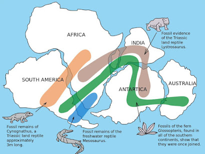
Maps of the Atlantic Ocean were produced, and people began to notice how well the continents seemed to fit together. However, since no one thought continents could move, this did not attract serious attention. In 1912, the geophysicist Alfred Wegener published a theory that 300 million years ago a single continent existed, and he named this ‘super-continent’ Pangaea. His theory suggested that today’s continents were formed from splitting of this giant mass. Wegener claimed his theory of continental drift was supported by the following evidence that these areas were once joined:


GEOLOGICAL evidence such as the near perfect fit of South America and west Africa as well as the fact that glaciation deposits of the same nature were found in South America, Antarctica and India. This could only occur if these masses of land were once connected.

BIOLOGICAL evidence including the fossil remains of a reptile that were found in both South America and southern Africa – it is unlikely that this reptile developed in both areas nor that it migrated across the Atlantic.

CLIMATOLOGICAL evidence of coal deposits that would have been formed in tropical climate conditions being found in places that do not have a tropical climate. E.g. Antarctica, N. America and UK.

How do we know the plates are moving?

Wegener’s theories couldn’t explain how continental movement takes place. From the 1940s onwards, evidence began to accumulate:

The mid-Atlantic ridge was discovered and studied alongside a similar feature in the Pacific Ocean. It was found to be 1000km wide, reaching heights of 2.5km and composed of volcanic rocks. Examination of the crust either side of the ridge revealed a paleomagnetic pattern – i.e. alternating polarity of rocks - suggesting that sea-floor spreading was occurring. The striped pattern, mirrored exactly on either side of a mid-oceanic ridge, suggests that the oceanic crust is slowly spreading away from this boundary. Very young rocks (less than 1 million years old) are found near the ridges and older rocks (over 200 million years old) are found near the continents. Sea-floor spreading implies that the Earth must be getting bigger – since this is obviously not the case, plates must be being destroyed somewhere. With this in mind, large oceanic trenches were discovered where large areas of ocean floor were being pulled downwards in subduction.

What is the mechanism behind the movement?

Since all plates move at their own rate, the forces acting on them must vary from plate to plate. It is therefore unlikely that any single agent moves the crust – movement must be controlled by a combination of forces:

1.           Convection Currents

Hot spots around the core of the Earth generate thermal convection currents within the asthenosphere, which cause magma to rise towards the crust and then spread before cooling and sinking. This circulation drives the movement of crustal plates. This is a continuous process, with new crust being formed along the line of constructive boundaries (divergent – plates moving away from each other) between plates and older crust being destroyed at destructive boundaries (convergent – plates move towards each other). Where plates move past each other parallel to a plate margin, there is no creation/destruction – at these conservative margins there is no subduction and no volcanic activity.


2.           Gravity (a more recent theory)

·       ‘RIDGE PUSH’ or gravitational sliding away from a spreading ocean ridge. Upwelling of fresh magma at constructive boundary creates a buoyancy effect resulting in the ocean ridge being at higher elevation off the ocean floor. Gravity acts down to slope of the elevated ridge, pulling the plate downwards and away from the ridge.

·       ‘SLAB PULL’ at destructive boundary. Movement is driven by the weight of cold, older, dense plate material sinking into the mantle which pulls the whole oceanic plate down. Frictional resistance as the plate is dragged down results in shallow and deep earthquakes at the subduction zone.  

Plates move in different directions, towards, alongside and away from each other. The direction they move in and whether they are continental plates or oceanic plates determines what happens in the plate boundary area. There are 4 types of plate boundaries:

       Divergent/constructive boundaries -- where new crust is generated as the plates pull away from each other. In oceanic areas, sea floor spreading creates a mid-ocean ridge whereas in continental areas, stretching and collapsing of crust creates rift valleys.

       Convergent boundaries -- where crust is destroyed as one plate dives under another or is crumpled upwards.

-          Destructive: oceanic & continental plates move towards each other. The denser oceanic plate is sub-ducted beneath the continental plate creating an ocean trench. Friction between plates often results in earthquakes. When this friction is combined with heat from mantle, the oceanic plate melts and the resulting hot magma rises, creating a volcano prone to violent eruptions.

-          Collision: two continental plates crash into each other and since densities are near identical, neither plate can be sub-ducted.  Instead, the force pushes plates together, again often resulting in earthquakes. The pressure causes rocks to bend and fold, creating mountains with no volcanic activity.

      Conservative/transform boundaries -- where crust is neither produced nor destroyed as the plates slide horizontally past each other. The sliding plates get stuck/jammed against each other so huge amounts of pressure build up. Eventually this pressure is released causing violent earthquakes and the plates move only millimetres. This kind of boundary is not typically associated with volcanic activity. The San Andreas fault is an example of a conservative boundary and it typically moves 33mm a year.

 Sources:

AQA Geography textbook for AS and A Level

https://www.nationalgeographic.org/media/plate-tectonics/#:~:text=The%20theory%20of%20plate%20tectonics,boundaries%20all%20over%20the%20planet. 


Monday, 3 August 2020

The Science Behind the Artificial Activation of Egg Cells

Briefly mentioned on our biology specification was the idea that human egg cells could be artificially activated to divide, without any input from sperm, essentially creating a source of embryonic stem cells whilst bypassing the ethical issues associated with such cells. That was as far as the textbook cared to explain so I went on a bit of a Google deep-dive to find out more about the process behind this.


Karl Swann is the Chair of Reproductive Cell Biology in the School of Medicine at Cardiff University. Swann’s team tricked egg cells into dividing by injecting them with an enzyme (phospholipase C-zeta) produced by sperm during natural fertilisation. Egg cells contain two sets of chromosomes and during natural fertilisation one of these is typically discarded within two hours; the team used a standard chemical treatment to prevent this resulting in a parthenogenetic embryo - one that contains no paternal chromosomes and instead has two sets from the mother - that appears to undergo the same changes as a natural embryo.

The activated eggs divide for around five days until they reach the blastocyst stage (at which they are around 100 cells strong) when they should in theory provide a source of stem cells. The embryos that are created from this process cannot develop into babies since no sperm was involved in the activation. Therefore, this sidesteps the ethical controversy around the destruction of potential human life, since genetically, these cells are not capable of life. The hope is that the pluripotency of these cells can be used to treat a range of diseases.

The enzyme used might also help women become pregnant through IVF. One IVF technique injects sperm directly into the eggs in a lab and then implants them into the uterus. Unsuccessful attempts occur when the embryos never begin dividing and it is thought this could be due to the sperm having defective PLC-zeta. Adding the enzyme artificially might start them dividing.

This isn’t the only method being trialled, in fact there are many routes to getting an ethical source of stem cells that are being looked into. For example, a team led by David Wininger grew parthenogenetic human blastocysts by stimulating eggs chemically and this approach involved triggering a calcium wave.

Another revolutionary and ethically optimal option would be the creation of embryos specifically for the purpose of isolating stem cells via ‘nuclear transfer’ or ‘therapeutic cloning’. This method involves the insertion of the nucleus of a somatic cell (any cell of a living organism other than the reproductive cells) into an enucleated unfertilised egg (one that has had the nucleus removed). Since the nuclear DNA of the cells is derived from a somatic cell of the patient intending to receive the transplant, the chances of tissue rejection are greatly reduced. The egg in this case is not fertilised. Instead it receives maternal and paternal genomes from the somatic cell nucleus. Since by some definitions an embryo is the result of fertilization of an egg by sperm, there is no absolute consensus that nuclear transfer gives rise to an embryo. Therefore, this is another way around the ethical issues of the embryonic stem cells having a right to life, since the artificially created ‘embryo’ wouldn’t produce a fetus if placed in the womb.


(An oocyte is a cell in an ovary which may undergo meiotic division to form an ovum. Autologous tissue is tissue that comes from the host itself - this relates to the reduced chance of rejection since the body recognises the new tissue as its own).

The ethical guidelines around this subject are also up for debate and it has previously been proposed that the criteria applied to organ donation could also be applied to the collection of embryonic stem cells from non-viable (essentially dead) human embryos produced during routine IVF procedures. It has been shown that such embryos do not continue normal embryonic development once in culture, yet most of them contain a substantial number of living cells on day 6 of culture, which suggests that non-viable embryos could provide reliable stem cell lines.


Sources:

https://www.newscientist.com/article/dn6733-zapped-human-eggs-divide-without-sperm/

https://academic.oup.com/humrep/article/18/4/672/596542

https://www.sciencedirect.com/science/article/abs/pii/S1472648310602702


Wednesday, 29 July 2020

The Science Behind Unique Fingerprints

I am loving the Infection, Immunity and Forensics topic that we are currently covering in Year 2 of A-Level Biology. The specification briefly talks about conventional methods of identifying a body, one of which is the use of fingerprints. This method is dependent upon the fact that all fingerprints are unique to an individual and never change throughout one’s lifetime. The global population stands at just under 7.6 billion people and the chance of your fingerprint being identical to someone else’s is 1 in 64 billion! So, what processes occur that mean every single fingerprint is different? How do we get our fingerprints?

Fingerprints are small ridges caused by folds in the epidermis of the skin; these ridges vary in length and width and can branch or join together to form distinctive patterns. They are even more unique than DNA – although identical twins mostly share the same DNA, they never have the same fingerprints. Each ridge contains pores that are attached to sweat glands under the skin. It’s because of this sweat that you leave finger marks on pretty much everything you touch. 

Unique fingerprints are the result multifactorial inheritance – i.e. affected by both your genes and the environment. The ridges on the tips of your fingers are frictional, and although small, they actually stick up above the rest of the skin. These friction ridges grow in different designs, as mentioned above, and if your parents have a certain pattern, you are likely to have it too; genes give the basic design of the finger ridge pattern, almost like laying fingerprint foundations. These genes also dictate how and when the skin grows. As a fetus develops in the womb, the dermis and epidermis of the skin grow together with friction ridges appearing where the two layers meet, and this is guided by genes. Since growth is entirely natural, and therefore not uniform, the layers grow at different speeds; when one layer grows faster than the other, it stretches and pulls cells in the other layer. In addition, a fetus’ fingers can rub against the inside of the womb. These tiny forces move the skin as it grows, with the cumulative effect being control over that direction of growing ridges which forms the unique fingerprint. Since everyone’s skin grows in slightly different environments, it is highly unlikely that your fingerprint will ever be identical to someone else’s.


Sources:

·       SNAB A2 Biology textbook

·       https://askdruniverse.wsu.edu/2020/02/07/people-different-fingerprints/

·       https://science.howstuffworks.com/fingerprinting1.htm

Tuesday, 28 July 2020

The Science Behind Sleep

You may be surprised to learn that on average we spend 26 years of our lives asleep. Moreover, we spend a further 7 years trying to get to sleep! But have you ever wondered what your body is actually doing while you’re asleep? Well read on and your questions will be answered.

It was once thought that during sleep, your body completely shuts down, both physically and mentally. Obviously, this cannot be the case since, while we sleep, we continue to breathe and carry out other processes such as cell repair that are vital for life. In fact, overnight your brain and body work hard to ensure that when you wake up, your body is ready for action. During sleep your brain repeatedly goes through a cycle of two different types of sleep, REM (rapid-eye-movement) and non-REM sleep. 


The first part of this cycle is non-REM sleep, which is broken down into four stages. Stage one comes between being awake and falling asleep and stage two is light sleep. This is when breathing and heart rate regulate and body temperature drops. Deep sleep makes up the third and fourth stages. You then cycle into REM sleep, the phase where dreaming occurs. The eyes move rapidly behind closed lids (hence the name REM) and your brain waves alter to be similar to those during the day when we are conscious and awake. While the rate of breathing increases, the body becomes temporarily paralysed as we dream. The cycle repeats itself, on average four to five times in one night, but with each cycle you spend less time in the deeper stages three and four of sleep and more time in REM sleep.

Have you ever heard people refer to their natural body clock? That they naturally wake up and fall asleep at the same time every day because their body has supposedly ‘set’ a built-in alarm?

According to research there are two main processes that actually promote this regulated sleep pattern: sleep drive and circadian rhythms.

Much like when it's nearing lunchtime and you can’t wait to dig into a massive slice of cake (no? just me then), your body craves sleep. As the day goes on, your desire for sleep builds, until it reaches a climax where you simply have to shut your eyes. Unlike hunger, where your body can’t force you to ingest food, it can put you to sleep, no matter the circumstances and you have no control over this. When you are exhausted your body even has the ability to engage in what’s called microsleep episodes of one or two seconds when your eyes are open. I have never been one to voluntarily take a nap, but it has been shown that napping for 30 minutes or more late into the day can derail your night’s sleep by decreasing your body’s sleep drive.

Your circadian rhythm is basically a 24-hour internal biological clock that is running in the background of your brain and cycles between sleepiness and alertness at regular intervals. A key function of this clock is responding to light cues, increasing production of the hormone melatonin at night, then switching it off when it senses light. Melatonin is a natural hormone that is produced by the pineal gland in your brain and acts on receptors in your body to encourage sleep.


So, why do we actually need sleep?

It has been proven that sleep significantly impacts upon brain function, notably its ability to accommodate inputs. Too little sleep leaves us unable to process things learnt during the day and reduces our ability to remember it in the future. Researchers also believe that sleep may promote the removal of waste products from brain cells. This process is still carried out while we are awake, just less efficiently. Health risks also rise when people consistently don’t get enough sleep with symptoms of depression, seizures and high blood pressure worsening. During sleep, our bodies try to conserve energy and hence sleep plays a key role in metabolism and people who chronically get fewer than six hours of sleep per night are more likely be obese and diabetic. Immunity is also compromised, increasing the likelihood of illness and infection.

References:

·       https://www.webmd.com/sleep-disorders/ss/slideshow-sleep-body-effects

· https://www.hopkinsmedicine.org/health/wellness-and-prevention/the-science-of-sleep-understanding-what-happens-when-you-sleep

·       https://www.sleepfoundation.org/articles/what-circadian-rhythm

·       https://www.nhs.uk/medicines/melatonin/

·       https://www.sciencedaily.com/releases/2020/04/200422091205.htm


Monday, 27 July 2020

The Science Behind 'that' Rain Smell

Disappointed as I was about having to cancel my end-of-a-weird-term garden party (taking place at a social distance, of course), I do love a good rainy day. I was out walking my dog when I smelt that distinctive odour of fresh rain… this got me thinking. Where does that all-too-familiar rain smell come from?


It would be frankly ridiculous to assume that water falling from the sky, the same H2O that comes out of my kitchen tap, has any kind of scent. And yet, a certain ‘earthy’ and fresh smell is evident in the air when it rains. The name of this smell is petrichor.

Definition: a pleasant smell that frequently accompanies the first rain after a long period of warm, dry weather.

This comes from the Greek word, meaning ‘the blood of stones’ and was coined by two Australian scientists in the 1960s, Isabel Bear and Richard Thomas, who were searching for the origin of the scent. Its sources were identified in two places, plants and bacteria, living in the soil.

The scientists found they could extract a yellowy oil from warm, dry rocks, clay and soil. This oil contained fatty acids from plants, namely palmitic acid and stearic acid, as well as other smaller compounds. Alone these compounds aren’t particularly odorous, but after some time in the soil, it was found they get broken down into smaller, much smellier molecules.

The other key contributor are bacteria in the soil called actinomycetes, tiny microorganisms that can be found in rural and urban areas as well as in marine environments. They decompose dead organic matter into simple chemical compounds which can then become nutrients for developing plants and other organisms. A by-product of this activity is geosmin, meaning ‘the smell of earth’, an organic compound that, when combined with the broken-down fatty acids, adds to the petrichor scent. Geosmin is a type of alcohol, and like most alcohol molecules, tends to have a strong scent, but the complex chemical structure of geosmin means the human nose can detect it at concentrations less than 10 parts per trillion – in context, around a teaspoon in 200 Olympic sized swimming pools!

So why is that we only associate the distinctive smell with rain? Why can’t we smell it all the time since it is always present in the soil?

The decomposition activity rate of actinomycetes slows considerably during a prolonged period of dryness. The air becomes more humid, or as many refer to it ‘muggy’, just before a rain event and the ground begins to moisten. This speeds up the activity rate of the bacteria, leading to increased production of that odorous geosmin. 


In 2015, research was carried out into how the smell actually reaches your nose. High-speed videos of water droplets hitting a porous surface, showed air bubbles getting trapped beneath the rain drop. Those air bubbles then burst, spewing tiny jets of water, effectively creating a natural aerosol of water from the raindrops and chemicals from the soil. These small water drops can travel on the wind much more easily than raindrops and hence carry the smell of petrichor a considerable distance both geographically across an area but also from the ground to our noses. Only a light rain will have this effect as heavy rain doesn’t create these bubbles and this scent eventually goes away after the rain has passed and the ground begins to dry.

P.S. for those equally disappointed about my lack of garden party fun, I rearranged to Friday when the weather is supposed to be delightful. Please cross your fingers and toes that this is the case!

References:

·       https://www.sciencenewsforstudents.org/article/scientists-say-petrichor

·       https://www.youtube.com/watch?v=2txpbrjnLiY&feature=emb_title

·       https://earthsky.org/earth/what-is-smell-of-rain-petrichor

Sunday, 14 June 2020

A Reading Recommendation: How We Live and Why We Die


I’m a BIG biology fan. Naturally when I came across this book – ‘How We Live and Why We Die’ – the complexity of the potential answers to these questions sparked my curious mind and I couldn’t wait to get started.


The way in which Lewis Wolpert shares his ideas in this book, not only demystifies the impossible nature of how our society of cells functions to give us the quality of life we know, but also highlights the volume of information left to uncover in this field of biology. Where exactly did the first cell come from? How do communicating nerve cells in the brain give us the capacity to think, feel and move? And do we actually die from ‘old age’ – or is it just the complications that come with age that are to blame? I was naïve to the idea that there is so much left to find out about cells and one day I hope to have an impact in the world of cells that helps to fill in some of the blanks. The possibility of new discoveries excites me!


If the secret lives of cells fascinates you and you are curious as to how exactly a single cell in your body works as part of society to keep you ticking along, then I can’t recommend this book enough. I am looking forward to doing further research into some of the topics covered in the book – as always I’ll share my findings so keep your eyes peeled!

Sunday, 7 June 2020

The Science Behind Sleep Paralysis

I recently listened to a podcast where the topic of ‘sleep paralysis’ came into conversation. Having never experienced the phenomena myself, but having many friends mention their brush with this paralysis, naturally I was curious to investigate further. A 2011 review showed that around 7.6% of the global population will experience at least one sleep paralysis episode in their lifetime, although there were higher rates recorded in students and psychiatric patients, particularly those with PTSD or a panic disorder.


So, what is sleep paralysis? From a non-scientific perspective, sleep paralysis locks a person in a temporary state between waking and dreaming, where they can't move but may experience odd hallucinations. People have been known to have a number of bizarre experiences from feeling pressure on their chest or a hand around their throat to a feeling of being removed from their frozen bodies, as if floating out of bed. This is sleep paralysis, a fairly common and diagnosable sleeping disorder.


Just why or how it happens isn’t fully understood yet. From a scientific standpoint, sleep paralysis is better understood now to be a neurological disorder, rather than a brush with the paranormal, that researchers believe arises from disrupted REM sleep. As a person falls asleep, their heart rate slows, muscle activity decreases, and brainwave activity slows in frequency. This is non-REM sleep. A normal sleep cycle involves a stage called rapid eye movement (REM); a unique phase of sleep in mammals and birds, distinguishable by random/rapid movement of the eyes, accompanied with the paralysis of voluntary muscles, and the tendency of the sleeper to dream vividly. It is during REM that about 90% of dreams occur. During REM sleep, we have three systems engaged: 

  1. Cortical activation – the brain is switched on creating the dream imagery.
  2. Sensory blockade – senses are blocked meaning no sight, no hearing and you stop feeling your body in the bed.
  3. Muscular paralysis – structure of the brain called ‘pons’ which sends signals to the spinal cord which paralyses the major muscle groups while you dream


Usually when you wake up, all three REM systems become disengaged at the same time. Sometimes, due to influencing factors such as jetlag, recreational drugs, sleep deprivation, or sometimes it just happens, two systems switch off while one stays engaged. When you wake up, cortical activation and sensory blockade are now switched off (we are awake, thinking hearing seeing etc.) while muscular paralysis stays on. The only body parts not paralysed during REM sleep and during this period are the respiratory system and eyes. Your eyes can look around and the respiratory system - because it is the only part not paralysed - is the only thing you can ‘feel’. Because of this, some people experience an intense pressure on their chest, pushing down and it feels like your breathing is very shallow because you can effectively feel the respiratory system in action. Your brain begins to think ‘well, if two of these systems are disengaged, but not three, maybe that means I should still be dreaming?’. In turn, the dreaming part of the brain switches back on; this happens whilst your eyes are open, so the imagery is referred to as a ‘psychotic hallucination’. So now suddenly you’re paralysed, experiencing pressure on your chest and you’re starting to hallucinate… and this is supposedly a normal and common disorder?!  Sometimes we hallucinate the last thing we were dreaming about.


Let’s take an example:


Say I’m dreaming about my dog (Alfie, a rather cute and fluffy spaniel in case you were curious). Suddenly my dog is sat at the foot of my bed. I love Alfie, but why is he in my room? This freaks me out and I feel scared; the brain responds to the emotional atmosphere of being freaked out and changes the hallucinations to fit. Now suddenly its not a cute fluffy spaniel, it’s a ferocious wolf bearing its teeth. Suddenly the wolf is linked to the feeling of pressure on the chest so now there’s a ferocious wolf pushing down on my chest and I’m having a full-on freak out.


That’s one possible explanation to the science behind sleep paralysis. Another is presented below by Daniel Dennis, a postdoctoral scholar in psychiatry:


"We know the amygdala is highly active in REM, which is important to fear and emotional memory. You have part of the brain actively responding to fear or something emotional, but nothing in the environment to account for that. So, the brain comes up with a solution to that paradox."


So how common is this? Up to as many as four out of every 10 people have some experience with sleep paralysis. A study in the Sleep Medicine journal of 185 patients diagnosed with sleep paralysis highlighted that around 58% sensed a non-human presence in the room with them and 22% saw a human in the room, typically a stranger.


What are the factors that cause sleep paralysis? Sleep experts believe that it may be partly genetic, but other factors include a history of trauma, poor physical health and sleep quality or substance abuse. The frequency and severity of episodes has also been linked to anxiety-like symptoms and sleep deprivation – this may explain why episodes come and go. They may coincide with periods of stress or anxiety.

Is it treatable?

Medication wise, no, there is no direct treatment for ‘sleep paralysis’. A routine change is usually advised to patients, including an improvement to sleep schedules and to maintain a better and more consistent bedtime routine. According to the National Health Society of the UK, in more extreme cases, patients may be prescribed a low dose of antidepressants. These medications may help lessen the symptoms of sleep paralysis by suppressing certain aspects of REM sleep. 

References:
https://www.youtube.com/watch?v=Hw7NFoKOIu4 
https://www.webmd.com/sleep-disorders/features/sleep-paralysis-demon-in-the-bedroom#1
https://www.livescience.com/50876-sleep-paralysis.html


Sunday, 17 May 2020

The Science Behind Meditation


Mindfulness is a type of meditation that involves focussing your attention on the present moment – for example, your body state and your breathing. It has been suggested in recent research studies that practising meditation can boost physical and mental health as well as cognitive abilities. Meditation is a way to build physiological and psychological resiliency, and so reduced stress is also a commonly reported side effect.

In our daily activity most of our minds wander to any worries or distractions we may have, but we can train ourselves, by building a mindful muscle, to be focussed and present. The key to this is strengthening the prefrontal cortex - the cerebral cortex covering the front part of the frontal lobe. This brain region has been implicated in planning complex cognitive behaviour, personality expression, decision making, and moderating social behaviour. This is like the conductor of an orchestra; by bringing our attention back to the body and present moment, the prefrontal cortex is activated – it’s forming new synapses and connections, getting thicker and stronger like a muscle.

Structures in the human brain


An electroencephalogram (EEG) is a test used to evaluate the electrical activity in the brain, since brain cells communicate with each other through electrical impulses. Synchronised signals on an EEG are brain waves; when you meditate, alpha and theta waves (types of brain wave) increase and activity in some parts of the brain decreases allowing a person to focus. The idea is that after a sustained period of mediation (i.e. 8 weeks of regular practice), the changes in brain activity experienced during meditation will have a lasting effect on the brain. According to doctors, a person will in theory be able concentrate better, make faster decisions and remember more information. Most extraordinary, the brain may even become more energy efficient; this happens because you’re performing better but the brain is using less energy, meaning a decrease in neural activity. You might be able to just as well in your job but exert less mental energy so when you are finished a task you are less tired.

Can mediation change the physical structure of your brain? Recent research form Harvard University showed that just 8 weeks of meditation could physically change the shape of the brain. Everything we do, from learning a new skill to a daily routine, affects the brain; repeated practice of something, in this case meditation, will strengthen certain connections in the brain and may cause changes in grey matter density. Grey matter contains most of the brain's neuronal cell bodies, and changes in density are caused by neurogenesis – the growth of new neurons in the brain. Harvard research has shown that for those who meditate, grey matter has increased in key areas such as the prefrontal cortex – i.e. the parts of the brain that help us focus, learn, inhibit impulses and regulate emotions.

In terms of physical health, can meditation change the body? Knowledge on this area is limited, but some research shows it may be good for our health, by reducing inflammation and stress hormones.

A major focus of research in the field of meditation is the question of whether this practice can slow down the aging process. Signs of aging are present in all cells, specifically in a part of every cell called the telomere – telomeres are the caps at the end of each strand of DNA that protect our chromosomes. Without this protective coating, DNA strands become damaged and our cells can't do their job efficient – effectively this degradation of cell function is ‘aging’. As we age these protective caps get shorter and shorter and the shorter these are, the faster the aging process. Comparison between telomeres of meditators and non-meditators, showed that the telomere length (a measure of immune cell aging) of meditators was more stable and didn’t shorten as rapidly. Other studies have measured telomerase, the enzyme that protects the telomeres – they have shown telomerase can go up and consequently telomere length can be better maintained for those in the mediation group.
Telomere


Psychologically, meditation also has been proven to have great benefits; for example, a number of people use mediation to manage depression. People who practise mindfulness on average show better mental health than 70% of the population and those with depression and anxiety have even larger gains. Data shows that meditation reduces depressive symptoms and it halves the rate of depressive relapse. Meditation can be an alternative to medication, and statistics show it to be as effective as antidepressants at preventing relapse. It works by breaking the cycle of depressive thoughts – notice the thought, let it go and bring focus back to the present.

So how does mindfulness train the brain? Through exercising the ‘attention’ muscle, a certain level of control can be exerted over the amygdala. The amygdala is a roughly almond-shaped mass of grey matter inside each cerebral hemisphere of the brain, which when activated is involved with the experiencing of emotions such as fear and anxiety - in depression or anxiety it becomes overactive. After mediation, other parts of the brain (including the prefrontal cortex and the hippocampus) appear more in control of the amygdala and those regions exert more regulation which dampens the effect of that anxiety or fear. However, most researchers are only looking at the positive effects – if you, for instance, have a psychotic disorder or severe anxiety, allowing yourself to completely focus on your mind may not be so beneficial. 


Reference

Sunday, 26 April 2020

A Reading Recommendation: A Briefer History of Time


I wouldn’t consider myself a massive physics fan and was more than happy to step away from the subject after finishing my GCSEs, having said this, I recently read Stephen Hawking’s ‘A Briefer History of Time’ at the recommendation of my fabulous maths teacher, since it ties in nicely with some of the themes covered in the mechanics modules of A-Level maths. I was sceptical to read it at first, thinking ‘This is Stephen Hawking, director of research in this field at the University of Cambridge – as if I’m going to understand a word’ but was pleasantly surprised to find the content manageable and soon found myself glued to every chapter!


Although, some of the topics covered when straight over my ‘non-physicsy’ head, certain elements literally blew my mind:


Disclaimer: THESE ARE NOT MY IDEAS! Everything mentioned below is lifted entirely from Stephen Hawking’s work.

  •          Firstly, the concept that there is no absolute space. To contextualise this, I will describe the example, utilised by Hawking in A Briefer History of Time. Imagine someone travelling on a train bounces a ping-pong ball straight up and down, with one second in between bounces. To this person the ball hasn’t moved – it has a displacement of 0. To an observer beside the track, the two bounces would be 40m apart since that is the distance the train itself has travelled in between bounces. Both views are equally acceptable, and one should not be favoured over the other since both observers have the right to consider themselves at rest.
  • Relativity of Distance
  • Secondly, the concept of there being no absolute standard of rest and therefore, no universal agreement on the speed of an object. Again, using the example from the book, we refer back to the ping pong ball. This time, the passenger on the train, hits the ball towards the front of the train with an observed speed of 10mph. To an observer on the track to ping pong ball is travelling at this 10mph relative to the train, plus the speed of the train relative to the platform. As with the theory of a lack of absolute space, both views are technically correct. Hawking poses the question; how do you define speed – relative to the train or relative to the Earth? With no absolute standard of rest, the ball cannot be assigned an absolute speed.
    Different Speeds
As a science and maths student, I am someone who likes a definitive answer (How far has the ball moved?! What speed is the ball moving at?!) and I consequently find myself both baffled and amazed by these ideas.


In conclusion, I found the book to give a really interesting and enjoyable introduction to the immense topic that is understanding our universe – so I would highly recommend it to those finding themselves curious to know more about this field. (There are loads more mind-boggling concepts like the ones above!)

Monday, 13 April 2020

The Science Behind Mood Rings


The mood ring was invented in 1975 by Joshua Reynolds, a marketing executive from New York City, and experienced fad popularity in the 70s, with the stone in the ring changing colour, supposedly according to the mood or emotional state of the wearer being a fascinating concept. Even today, they are still around, with mood rings, necklaces and bracelets all holding a permanent spot in the jewellery market. 

Mood ring colour chart
Of course, the mood rings cannot relay your actual emotional state with any degree of accuracy (since they possess no psychic abilities!) but the science behind the colour changing stone can be scientifically explained.


The ‘gem’ or ‘stone’ in the ring is really a hollow shell or quartz or glass encasing thermochromic (undergoes a reversible change in colour when heated or cooled) liquid crystals. The metal band of the mood rings conducts heat from the finger to the liquid crystal, which changes colour in response to the temperature of the skin. There are several natural and synthetic crystals that possess these temperature-sensitive properties, but the most common organic polymer used to make the crystals is based on cholesterol. More energy is available to the crystals as the ring becomes warmer; the molecules absorb the energy and twist which affects the way light passes through them. This means that the wavelengths of light that are absorbed or reflected by the ‘stone’ are affected.

The crystals are calibrated to reflect colours that are associated with certain moods; for instance, they are manipulated to reflect and therefore display a pleasing blue/green colour at the average resting peripheral temperature of 37.5ᵒc – implying the wearer is calm and relaxed. External body temperature increases in response to emotions of passion and happiness, and the crystals twist to reflect more blue light as they respond to the thermal change.  This is called the nematic phase; it is characterised by the rod-shaped crystal molecules pointing in the same directions but without any specific order. When the wearer is excited or stressed, blood flow is directed away from the skin to vital organs which require the excess glucose and oxygen to function in this state. For instance, when in a stressful situation the body goes into a ‘fight or flight’ response that triggers numerous hormonal changes. In the heart for example, the extra adrenaline binds the heart causing it to beat faster and pump a higher volume of blood quickly. This diversion of blood flow leaves the fingers cooler, causing the crystals to twist in the opposite direction, to reflect more yellow light, creating the red and amber colours. This is the cooler smectic phase, when the crystal components are aligned and display a degree of order.  

So, although mood rings do not have the psychic power to reflect what mood you’re in, the composition of the crystals does give the ‘gem’ the ability to respond to changes in body temperature… which to a certain extent can be related back to the emotional state of the wearer. Kind of cool really! 

References:
https://www.thoughtco.com/mood-rings-thermochromic-liquid-crystals-608013 

Friday, 3 April 2020

The Science Behind 3D Printing Organs


  Currently, there are hundreds of thousands of people on transplant lists all over the world, waiting for critical organs like kidneys, hearts, and livers that could save their lives. I was shocked to hear in a recent assembly that last year, over 400 people in the UK died waiting for a vital organ transplant and a further 777 people were removed form the waiting list due to deteriorating health that meant a transplant was no longer a viable option. As it stands, in the UK, 5693 patients are waiting for a transplant and statistics show that in the USA every 10 minutes another person is added to the transplant waiting list, since unfortunately, there aren’t nearly enough donor organs available to fill the growing demand.


This got me thinking; in this crazy and unprecedented world of modern research and development, surely there is a solution to this problem?

And yes, there is.

  Bioprinting is a branch of regenerative medicine currently under development that has the potential to create brand new, customised organs from scratch. Like modern 3D printing, bioprinting is a technique that deposits layers of material on top of each other to construct a 3D object one slice at a time, but instead of starting with plastic or ceramic based inks, a 3D printer for tissues and organs uses bioink.

  The main component of many bioinks are water-rich molecules called hydrogels. Millions of living cells are mixed into these, as well as numerous chemicals encouraging processes such as cell communication and growth. Engineering an organ or tissue is dependent upon having the right kinds of cells. In some cases, cells are isolated from a small tissue sample the size of a postage stamp. They are then mixed with growth factors and multiplied in the lab. The cells rapidly multiply in quantity so that, in about 6 weeks, a layer one cell thick could theoretically cover a football pitch. For cell types that cannot be substantially grown outside the body (e.g. heart, nerve, liver and pancreas cells), stem cells may be an option because of their pluripotency – the ability to become multiple cell types. Some inks contain just a single type of cell, whilst others combine several different kinds of cells, enabling scientists to create more complex structures.

  There are several printing techniques utilised in this field, the most popular being extrusion-based bioprinting. In this, bioink gets loaded into a printing chamber and pushed through a round nozzle attached to a printhead. It emerges from a tiny nozzle to produce a continuous fibre, the thickness of a human fingernail. 

  A computerised image guides the placement of the strands, either onto a flat surface or into a liquid bath that’ll help hold the structure in place until it stabilizes. After printing, some bioinks will stiffen immediately; others need UV light or an additional chemical or physical process to stabilise the structure. A successful printing process means that the cells in the synthetic tissue will begin to behave the same way cells do in real tissue: signalling to each other, exchanging nutrients, and multiplying.

  Most bioprinting uses a scaffold to hold cells in place. And once cells are ‘coaxed’ to a certain level, they begin to self-organise and assemble and then the scaffold can be removed. Dr Nakayama, a doctor and chairman of Saga University’s Regenerative Medicine and Biomedical Engineering department, has been developing a way to create 3D tissue without the need for scaffolds. Instead, he mounts small spheres on a fine array of needles called a kenzan and is now preparing the first human trial to implant dialysis tubes made entirely from a patient’s own skin cells. 

3D bioprinting using a kenzen rather than scaffolds
  The technology in this field already exists to print simple structures like lung tissue, skin, cartilage as well as miniature, semi-functional versions of solid organs, including kidneys livers and even hearts. Researchers have successfully used 15 different applications of cell/tissue therapy technologies in human patients - including a bioprinted bladder that was successfully implanted into 10-year-old Luke Massella in 2001.

  Luke Massella was born with a condition known as Spina Bifida, which left a gap in his spine. He is one of about 10 people alive walking around with a replacement bladder that has been grown from his own cells. At age 10, a malfunctioning bladder caused his kidneys to fail, and Luke faced a lifetime of dialysis treatment, that would have severely inhibited him from living a normal life. An enterprising surgeon, Dr Anthony Atala at Boston Children's Hospital, took a small piece of Luke's bladder, and over two months grew a new one in the lab, then in a 14-hour surgical procedure he replaced the defective bladder with the new one. In using the patient’s own cells this way, the issue of organ rejection (when the body’s immune system attacks transplanted cells from another organism) was minimised. Luke hasn’t had to have any surgery since and now, at 27, lives without complication.
Scientists at Wake Forest IRM using bioprinting to develop a replacement bladder.
  However, replicating the complex biochemical environment of a major organ is a harder feat, with so many more cells per centimetre. One of the biggest challenges is how to supply oxygen and nutrients to all the cells in a full-size organ and so the greatest successes so far have been with structures that are flat or hollow. To try and overcome this issue, researchers are working to incorporate blood vessels into the printed organs.  

  Ultimately, bioprinting organs from people's own cells will solve the huge lack of supply in organs for transplant and eliminate the need for anti-rejection immunosuppressant drugs. Specialist printers could even reproduce cancers tumours, giving doctors the chance to test treatments on specific patients. Bioprinters also provide a way of testing small quantities of fluid to test if a new antibiotic would work for a specific patient - this could help tackle the growing and serious problem of antimicrobial resistance.

  The potential to use bioprinting to save lives and advance our knowledge of organ function is huge. This kind of technology also opens up the possibility of augmentations similar to those seen in the classic science-fiction and superhero films, such as the printing of tissue with embedded electronics or the engineering of organs that exceed current human capability. Could we give ourselves futuristic qualities such as unburnable skin? And to what extent might we extend human life by printing and replacing our own organs when they start to fail with age?


References:
https://www.bbc.co.uk/news/business-45470799 https://www.ted.com/talks/taneka_jones_how_to_3d_print_human_tissue/transcript?language=en 
https://school.wakehealth.edu/Research/Institutes-and-Centers/Wake-Forest-Institute-for-Regenerative-Medicine/Research/ABCs-of-Organ-Engineering#Learn About the Steps Involved in Engineering Tissue and Organs